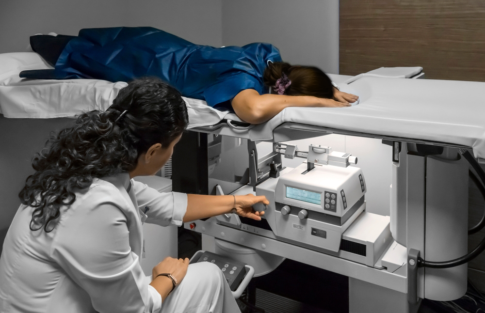
Magnetic Resonance Imaging or MRI-guided breast biopsy is the process of removing a small sample of breast tissue. It is the best procedure to determine the suspicious area for breast cancer. It uses.....
Magnetic Resonance Imaging or MRI-guided breast biopsy is the process of removing a small sample of breast tissue. It is the best procedure to determine the suspicious area for breast cancer. It uses powerful magnetic fields, radio waves; and a computer is used to help in locating a breast abnormality, and for the removing of a tissue sample with a needle for examination under a high-quality microscope. A magnetic resonance imaging-guided breast biopsy is widely helpful when MR imaging process indicates any kind of breast abnormality. The surgery is usually completed within 45 minutes, and the recovery involves no excessive bleeding or pain.
Vacuum-assisted breast biopsy is the best way to begin the treatment for breast cancer. Other conditions it may be used in are:
-
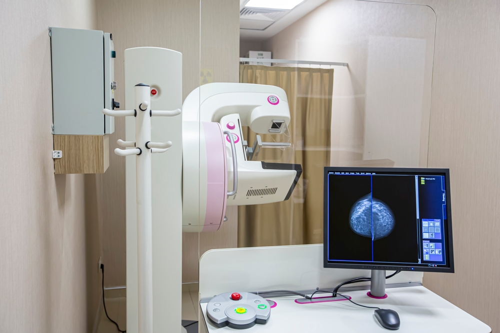
To detect the area of abnormal tissue change
-
Analyse suspicious mass not identified by other imaging procedures
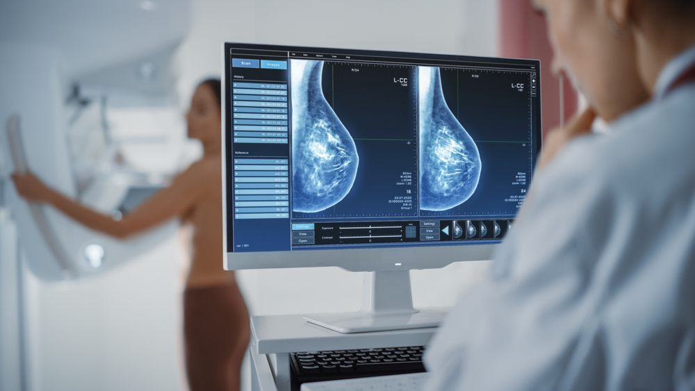
Preparing for the procedure
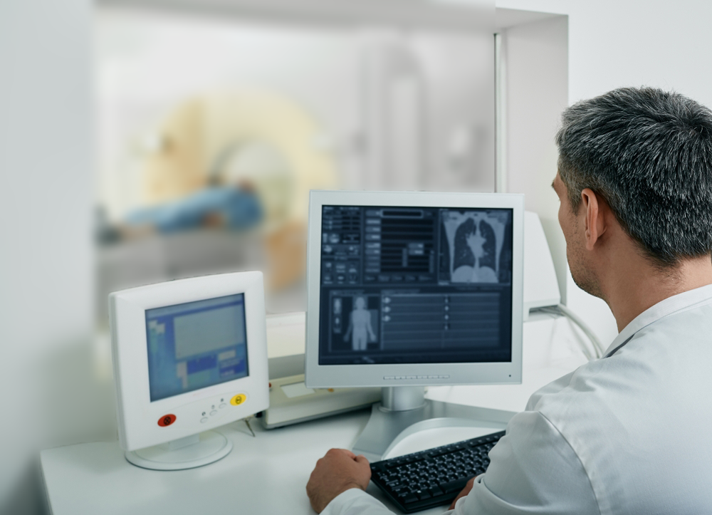
- The patient will be asked to change into a loosely fitted gown provided by Medanta.
- Before the procedure, the doctor should know about any allergies that the patient might be suffering from; especially the ones related to anaesthesia.
- The patient might have to undergo an MRI or CT scan before the procedure.
- The patient also has to inform about metallic implants and must take off entire metallic object if any.
During the treatment
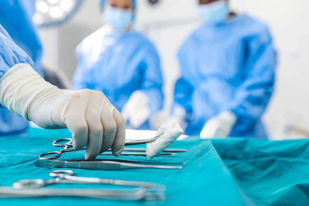
There is a need of an intravenous (IV) line that is inserted into a vein in the hand, and a contrast material gadolinium will be applied to improve the visibility of internal body structures. The breast will slowly compress between two compression plates, one of that would be marked with grid structure. The position of the lesion on the grid is measured by the radiologist with the help of the computer software and calculates the position and depth of the needle position. A localised anaesthetic is injected into the breast for the purpose of making the breast numb. Then, radiologist inserts and advances the needle to the area of the abnormality and MRI is performed to identify its position. Based on the method of MRI unit being used, the patient may remain in place or be taken away from the centre of the MRI scanner.
After the procedure
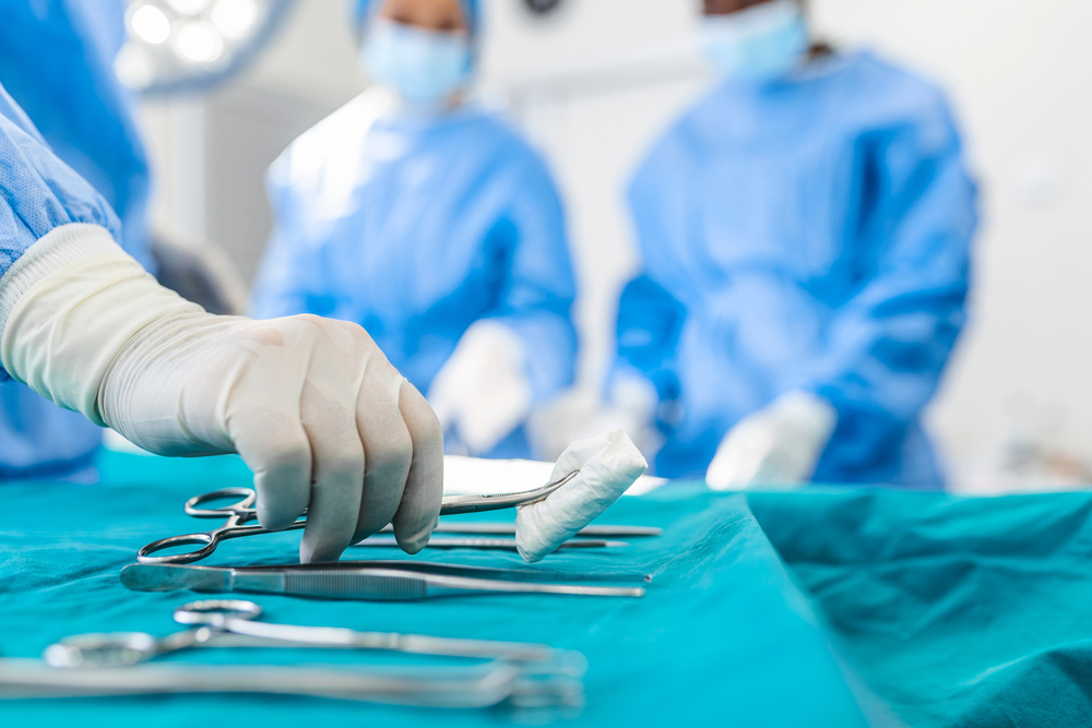
- After the procedure, the extracted lump will be sent to the laboratory for examination. The patient is usually allowed to go home after the procedure.
- If the patient experiences any side-effects or nausea, the same should be conveyed to the doctor immediately.
Vacuum breast biopsy treatment is considered to be the best for detection of breast cancer. The technology has some amazing benefits, but it also a few risks, and those are stated below:
-
The process is less invasive than a surgical biopsy. Does not involve exposure to ionising radiation. The breast biopsy that uses a core needle is safe and accurate. Causes less damage to tissue, takes less time and is less costly. Patients can quickly resume their usual activities, as the recovery time is fast.

-
The risk of bleeding or blood clots at the site of the biopsy. Increased risk of infection. Altered breast appearance. Bruising or swelling

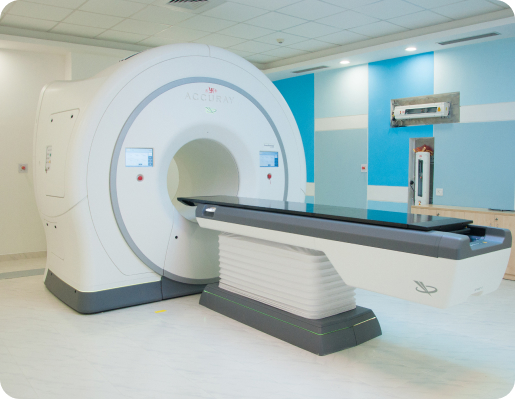
• The technology delivers quality samples from the most difficult areas within the breast.
• The sampling can be either automated or manual. The automated sampling enables support and time for the patient.
• Medanta’s MRI-guided breast biopsy surgery system includes Bard Encor Breast Biopsy technology whose speed and efficiency is unmatchable.
• There are two modes in the technology, dense and thin tissue modes that help the surgeon adjust in accordance with tissue density.
• The latest technology of vacuum-assisted breast biopsy surgery also provides versatile vacuum options for the fluid accumulation at the biopsy site.
